
Written By: Dr. Stephanie Mulick, O.D.
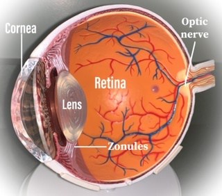
To understand diseases that affect the eye it is important to have a basic understanding of ocular anatomy. The eyeball consists of an anterior segment including the cornea, iris and lens and a posterior segment including the vitreous humor, the retina and the optic nerve.
Light enters our eye through the clear cornea, then the pupil and then through the crystalline lens at which point it passes through the vitreous body until it reaches the retina where it travels through our optic nerve and visual pathway to our brain where it is interpreted as what we see.
When it comes to visual acuity or our sight every structure in the eye needs to be healthy and functioning properly.
Sclera and Conjunctiva
The conjunctiva is the transparent (clear) membrane that covers the sclera (white of the eye) and the inside of the eyelids.
The sclera is comprised of collagen providing structure and protection to the eyeball. It is continuous with the clear cornea.
Conditions that affect the sclera and conjunctiva include:
- Pinguecula and pterygium (click here for more information)
- Conjunctivitis or pinkeye (to learn more click here)
- Cancer
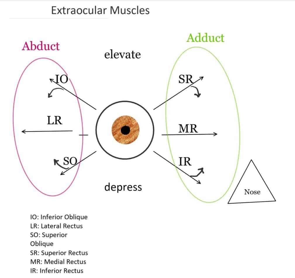
Extraocular Muscles
There are 6 extraocular muscles that control our eye movements and 1 muscle to control the superior eyelid.
The levator palpebrae superioris aids in lifting our eyelids.
There are four recti muscles: Superior rectus, Inferior rectus, Medial rectus and Lateral rectus.
The superior rectus aids in adduction (movement toward the nose) as well as rotation and upward movement of the eye.
The inferior rectus aids in abduction (movement away from the nose) as well as rotation and downward movement of the eye.
The Medial rectus aids in adduction (movement toward the nose) of the eye.
The Lateral rectus aids in abduction (movement away from the nose).
There are two oblique muscles, the superior and inferior oblique muscles.
The superior oblique abducts, rotates and depresses
The inferior oblique adducts, rotates and elevates.
Cornea
As stated earlier light enters our eyes through the clear cornea, if the cornea is not clear or healthy then light will be scattered as if looking through frosted glass.
There are several conditions that can affect the clarity of the cornea. (This is a list of some common corneal conditions)
- Dry Eye Syndrome (to learn more click here)
- Pterygium (Carnosidad): A non-cancerous vascular (containing blood vessels) growth that progressively moves from the sclera (white of the eye) to the cornea, eventually can cover the pupil (where light enters the eye) causing a decrease in vision. A pterygium can be surgically removed. They are primarily caused by sun (UV) exposure and sometimes associated with dry eye.
- Corneal inflammation
- Inherited or congenital (present from birth) corneal diseases
- Acquired age-related corneal degenerations
- Over wearing contact lenses
- Corneal abrasions or trauma
- Conjunctivitis or infection of the cornea
- Corneal tumor
Anterior Chamber
The space between the cornea and the iris is termed the anterior chamber. The anterior chamber is filled with a clear fluid called the aqueous humour which provides nutrients to the eye. The aqueous humour is constantly being produced by the ciliary muscle at the base of the iris and drained by the canal of Schlemn at the angle of the eye. The ciliary muscle of the eye is responsible for accommodation/focus and regulating the flow of the aqueous humor.
The part of the eye where the cornea and iris meet forms an angle. This angle is important for aqueous outflow which aids in regulating the intraocular pressure (IOP) of the eye. When the angle of the eye is narrow it can cause the aqueous humour to drain slowly and cause an increase in intraocular pressure (IOP). When the IOP becomes high it can cause damage to the optic nerve (Glaucoma). Narrow-angle glaucoma is a result of high IOP from a narrow-angle.
Inflammation, trauma or infection can affect the anterior chamber of the eye leading to decreased vision or eye pain, which would require treatment by an eye doctor.
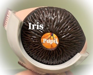
Iris and Pupil
The Iris is the colored part of the eye, it is in the shape of a doughnut with a hole in the center. The hole of the iris is the pupil where light enters into the eye. The pupil can dilate (enlarge) or constrict (become smaller) depending on the amount of light entering the eye. With more light, the pupil constricts. The pupil also constricts when we look up close to read.
Lens
Typically, the crystalline lens is an avascular transparent structure enclosed by a thin capsular membrane. The lens continues to grow throughout life. Eventually leading to presbyopia, loss of accommodation/focus, time for some reading glasses.
A cataract occurs when the crystalline lens of the eye becomes cloudy or opaque causing light to scatter or blur when it passes through similarly to frosted glass. With time this opacity will cause progressive blurry vision and if left untreated eventually visual impairment.
Here is a link for my post on Cataracts
Vitreous Humour
The vitreous humor is composed of a transparent gel-like substance that fills the space in the eyeball between the lens and the retina, this helps to maintain the structure of the eye by providing shock absorption and support to the retina. The vitreous humor must be clear so that light can pass through it. According to a Caltech thesis, the vitreous humor is ~ 99% water and 0.9% salts (and) 0.1% divided between protein and polysaccharide components.”
The vitreous is enclosed in a clear membrane called the hyaloid membrane, which is attached to the retina in several areas. As we age, the vitreous humor becomes more liquid, a process termed syneresis. Syneresis of the vitreous humor can cause it to shrink and collapse as well as pull away from the retina. This process can cause us to perceive floaters or wispy shadows in our vision and sometimes flashes of lights, due to pulling on the retina. The separation of the vitreous humor from the retina is called a Posterior Vitreous Detachment (PVD).
Here is a link to my post on Flashes and Floaters
Retina
The retina is the sensory tissue that lines the inside of our eyes. The retina is made up of photoreceptors (rods and cones). Rods are responsible for nighttime vision and cones aid in daytime vision. The area of the retina termed the macula has the maximum density of cones which contribute to fine detailed visual resolution.
Here is a link to my post on Macular Degeneration
Retinal cells transmit light signals to the optic nerve then through the visual pathway to the Cerebral cortex of our brain (Visual Cortex).
Optic Nerve
The optic nerve is also known as cranial nerve II. As stated above the optic nerve is necessary for the transmission of visual input to our brain. Several conditions can affect the optic nerve such as glaucoma, optic nerve inflammation, trauma and optic nerve atrophy.
Click here to learn more about Glaucoma.
Conclusion
The eyeball is a complex organ and each part is vital for visual clarity. Even if you have great vision, an annual eye exam is necessary to evaluate all aspects of the eye to ensure clear vision and healthy ocular anatomy throughout life.
hello there and thanks in your info – I have certainly picked up something new from right here. I did alternatively experience some technical points the usage of this web site, as I skilled to reload the website a lot of times previous to I may just get it to load correctly. I have been thinking about if your web host is OK? Now not that I’m complaining, however sluggish loading cases instances will often impact your placement in google and could injury your high quality score if advertising and ***********|advertising|advertising|advertising and *********** with Adwords. Anyway I’m adding this RSS to my email and can look out for a lot more of your respective fascinating content. Make sure you replace this once more very soon..
Please let me know if you’re looking for a writer for your site. You have some really great posts and I believe I would be a good asset. If you ever want to take some of the load off, I’d really like to write some material for your blog in exchange for a link back to mine. Please shoot me an e-mail if interested. Regards!
I’ve observed that in the world the present moment, video games will be the latest fad with children of all ages. Periodically it may be not possible to drag young kids away from the games. If you want the best of both worlds, there are numerous educational games for kids. Interesting post.
I think this is one of the most significant info for me. And i’m glad reading your article. But wanna remark on few general things, The site style is wonderful, the articles is really nice : D. Good job, cheers
Buy Private Proxies: Private and nameless, Best quality proxies, Indefinite bandwith, 1000 mb/s superspeed, 99,9 uptime, USA place proxies – https://DreamProxies.com